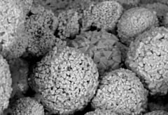
The time of tests: scanning electron microscopy and x-ray diffraction
In this third and final part of my introduction to the wonders of petrography I am going to briefly cover the techniques of scanning electron microscopy and x-ray diffraction.
Both these techniques provide powerful information that can be used to confirm or enhance petrographic examination, although they also come with potentially high costs and a range of limitations.
It is rare to really require these techniques, but they must always remain a potential consideration.
Scanning electron microscopy (SEM) involves firing a beam of electrons at (in this case) a piece of stone. The electrons can bounce back in different ways to create what is known as a back-scatter image, heavier elements usually bouncing back the most electrons and thus appearing brighter in the resolved images.
Some electrons are absorbed by the atoms at or near the surface, which leads to the release of secondary electrons from these atoms with properties that allow the nature of the atoms to be determined.
Because of the small size of the electrons, features down to a nanometer (a millionth of a millimetre) can be resolved.
The electron beam also causes atoms to release x-rays that can be detected and used to provide estimates of atomic composition. There is an awful lot of science involved.
SEM often suffers from not being able to see the wood for the trees because of the small size of the samples coupled with the high magnification that may skew the findings.
The lack of good contrast also makes spotting subtle variations sometimes very difficult. On the positive side, analysis can be used to confirm mineral types and assess changes in mineral concentrations. The technique is excellent for looking at clay minerals that are difficult to resolve optically, though x-ray diffraction may be preferred for assessing their potential performance.
X-ray diffraction (XRD) involves firing x-rays at a sample that is most commonly crushed up into a fine powder. The wavelengths of x-rays is roughly the same size as atoms, which they interact with and diffract in ways that are unique for each crystalline material. An x-ray diffraction fingerprint is produced by every mineral, which can be used to detect its presence.
The powder diffraction technique may be a little crude and not particularly good at identifying minerals present in proportions of less than around 1% of the sample, though different types of preparation improve the accuracy. Several traces may need to be run to provide a relative degree of quantities of minerals, often referred to as semi-quantitative XRD.
Where XRD comes into its own is with clay minerals, particularly those with potential swelling properties.
In many instances it is swelling clay minerals that lead to the decay of stone by making it unsound, often reducing quality or providing weak points for attack mechanisms to exploit.
By adding a chemical (typically glycol) to make the clay minerals swell, the amount of clay minerals with a potential for swelling can be determined and with it the relative level of potential unsoundness.
XRD is also useful for assessing roofing slates where the minerals are often too fine to be confidently identified using the optical microscope.
Both SEM and XRD work best in combination with optical microscopy. The combined findings provide very powerful evidence upon which quality conclusions can be reached and recommendations made.
It is worth getting to grips with these techniques to understand when and why you should use them. Not using them might be a lot more expensive than using them in the long term.

